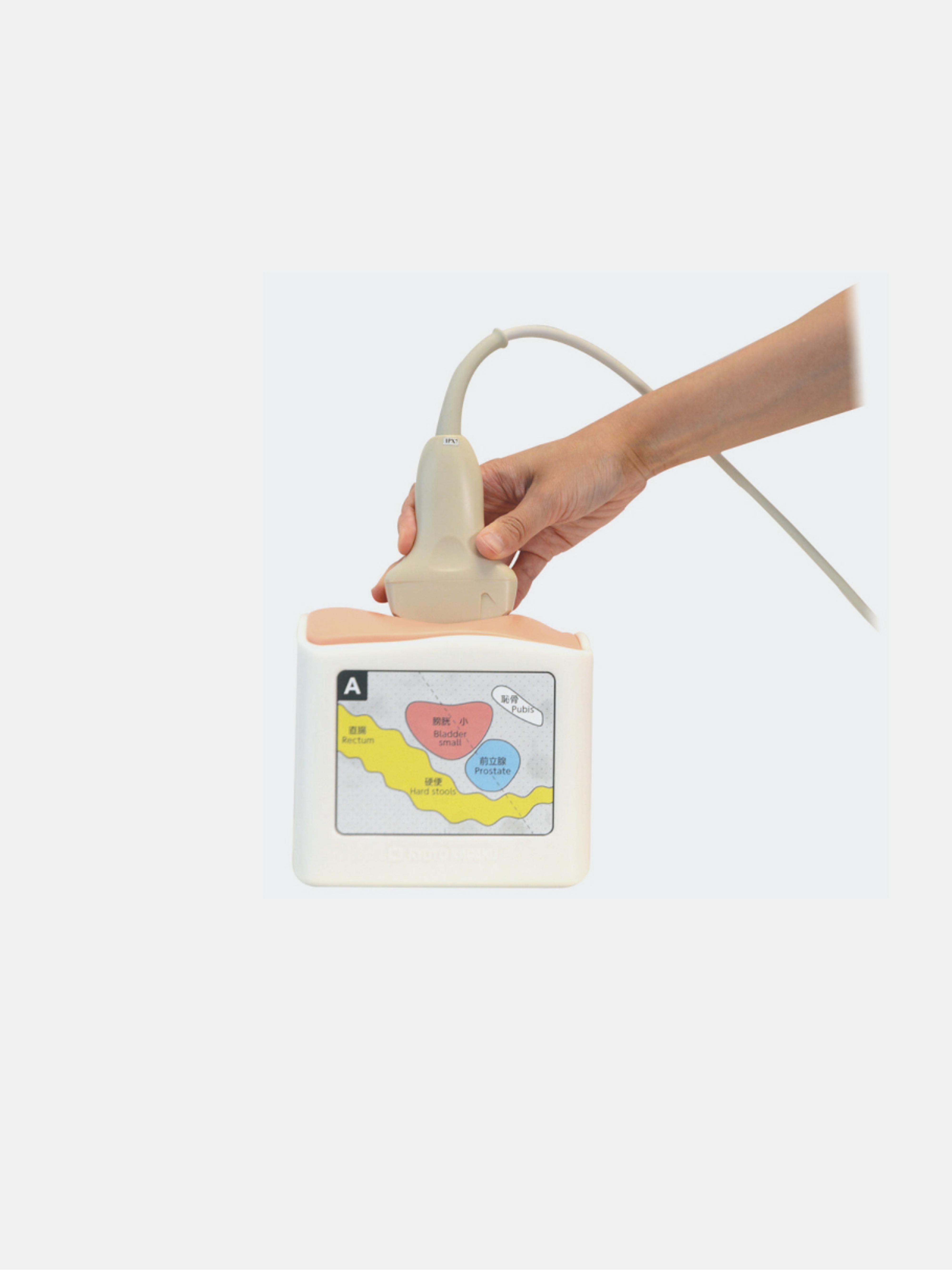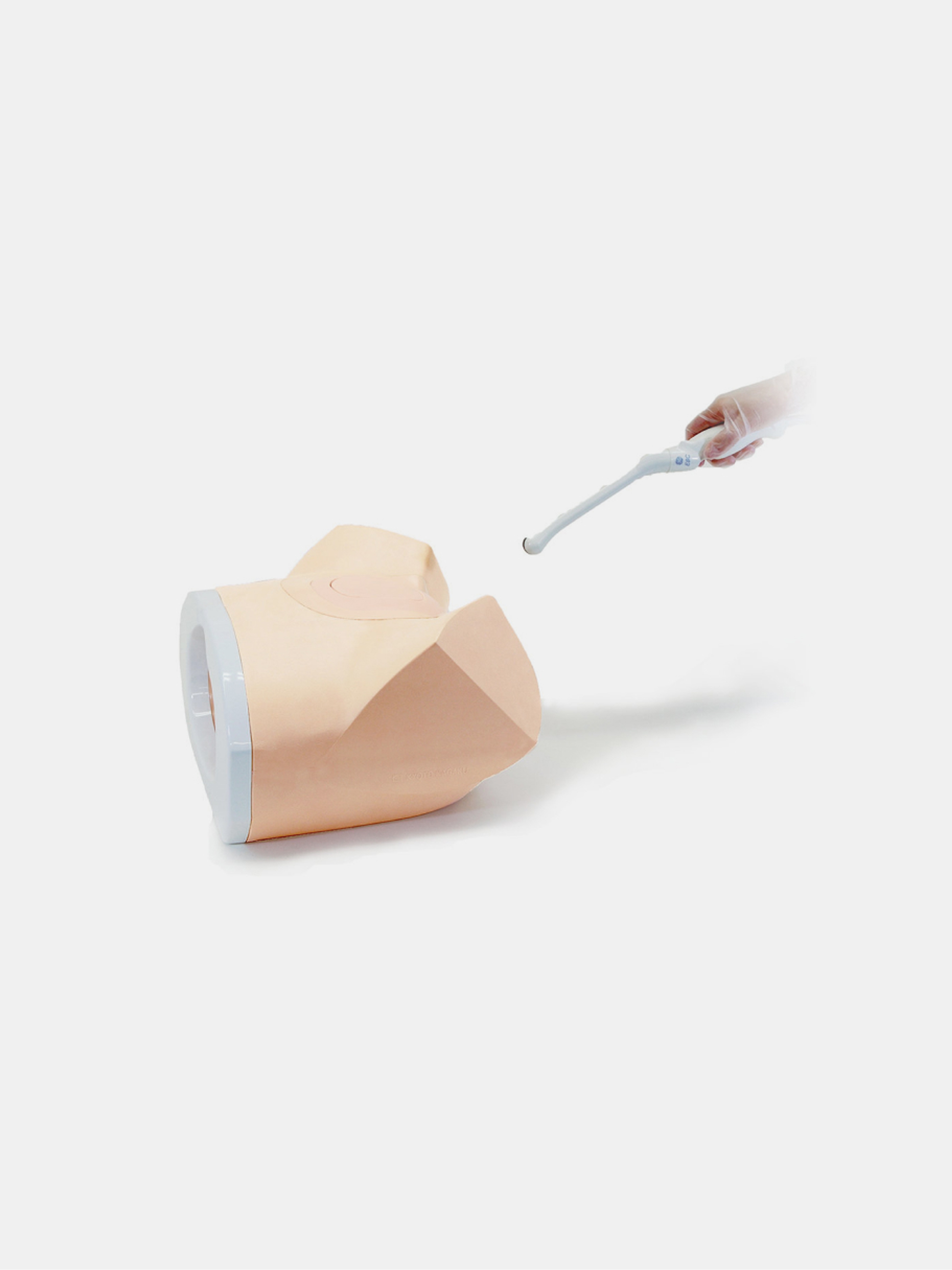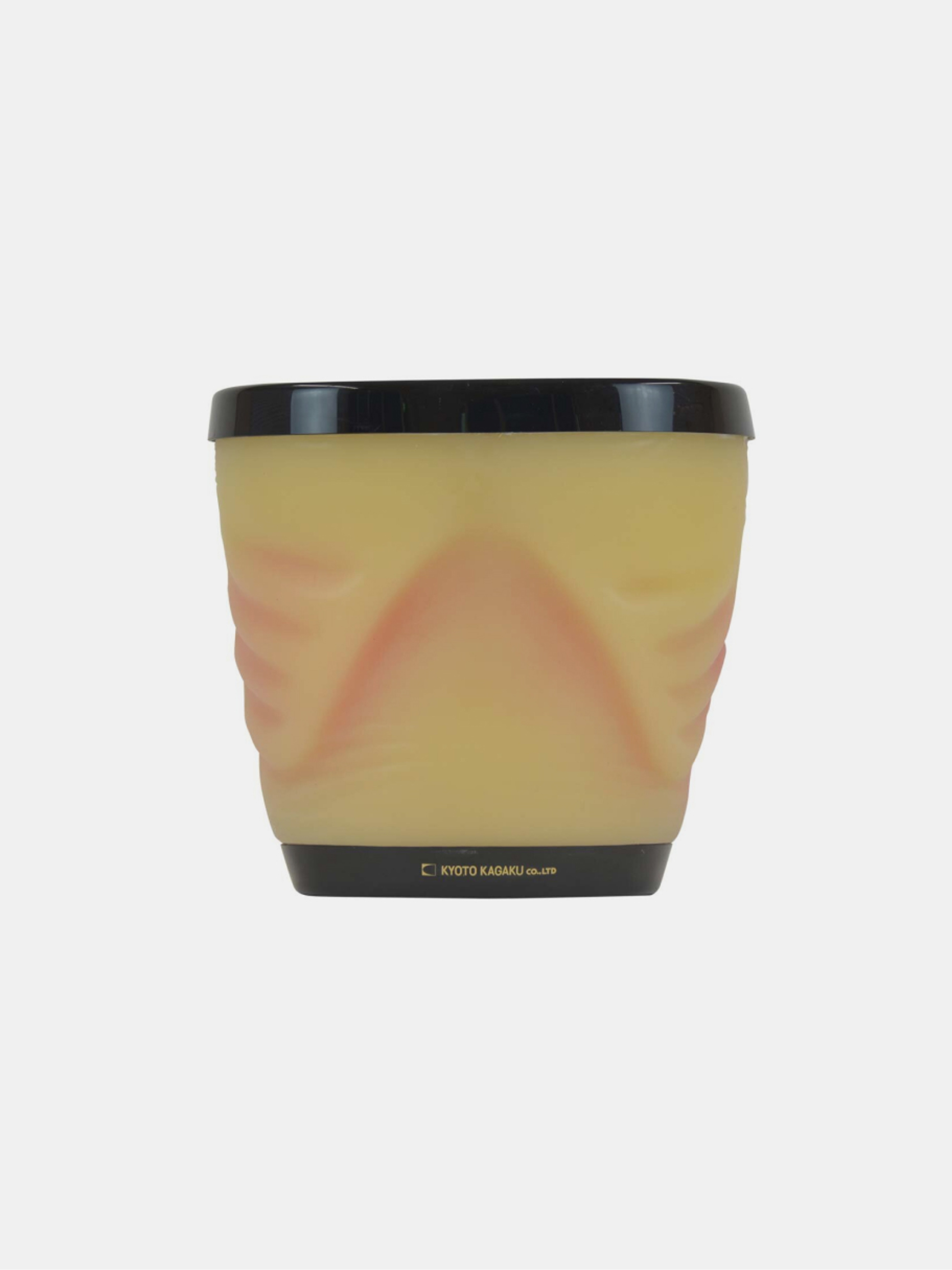
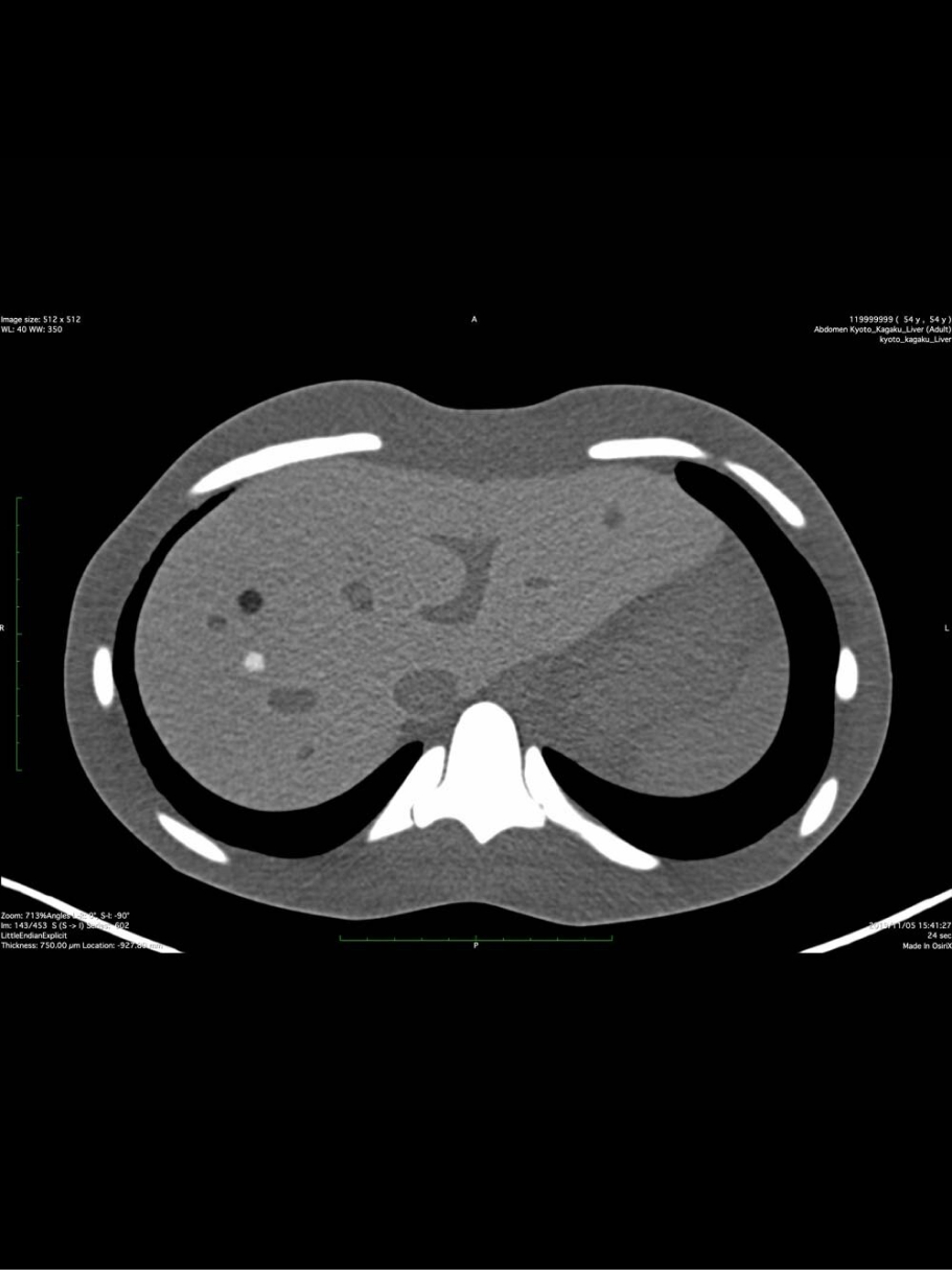
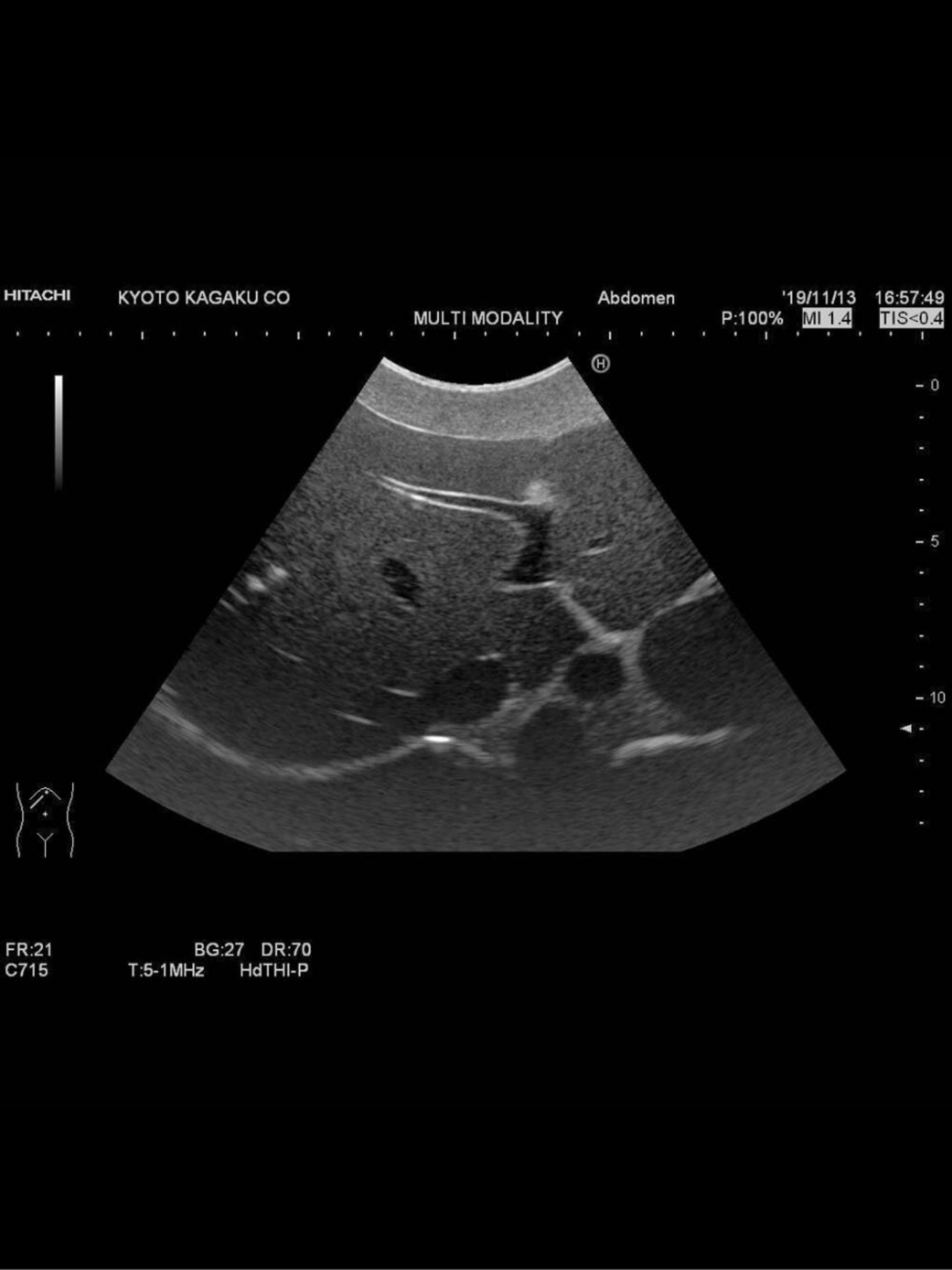
The dual-modality abdomen phantom (CT / Ultrasound) is designed for fusion imaging training and clinical research across CT and ultrasound modalities.
It enables fusion image generation, surgical and radiotherapy planning, and anatomical and vascular mapping under realistic imaging conditions.
The phantom accurately reproduces human abdominal anatomy, including the liver, biliary tract, spleen, kidneys, and major vascular structures such as the aorta, vena cava, celiac artery and branches, portal vein system, and renal vessels.
It also includes multiple pathological lesions for imaging evaluation:
- hepatic cystic and solid lesions
- gallbladder and bile duct stones
- pancreatic tumors (one invading the portal vein)
- splenic and renal lesions
- left adrenal tumor
It is ideal for fusion imaging studies, lesion localization, treatment planning, and biopsy mapping (no invasive puncture possible).
Specifications
- Applications: CT/Ultrasound fusion imaging, surgical and radiotherapy planning, anatomical mapping
- Included anatomy: liver, biliary system, spleen, kidneys, detailed vascular structures
- Simulated pathologies: hepatic lesions, biliary stones, pancreatic tumors, splenic and renal lesions, adrenal tumor
- Dimensions: 29 × 19 × 31 cm
- Weight: 12 kg
- Included accessories: positioning pillow, talcum powder, storage case, instruction manual
- Limitation: invasive procedures (biopsy or puncture) not possible
- Material: CT and ultrasound compatible tissue-equivalent material
Models
- 41952-000 - Dual Modality Human Abdomen Phantom (CT, Ultrasound) - US-22