Get in Touch
CT Whole Body Phantom "PBU-60"
The CT Whole Body Phantom PBU-60 is a realistic anthropomorphic model designed for radiology training and CT imaging optimization
0
Get in Touch
16
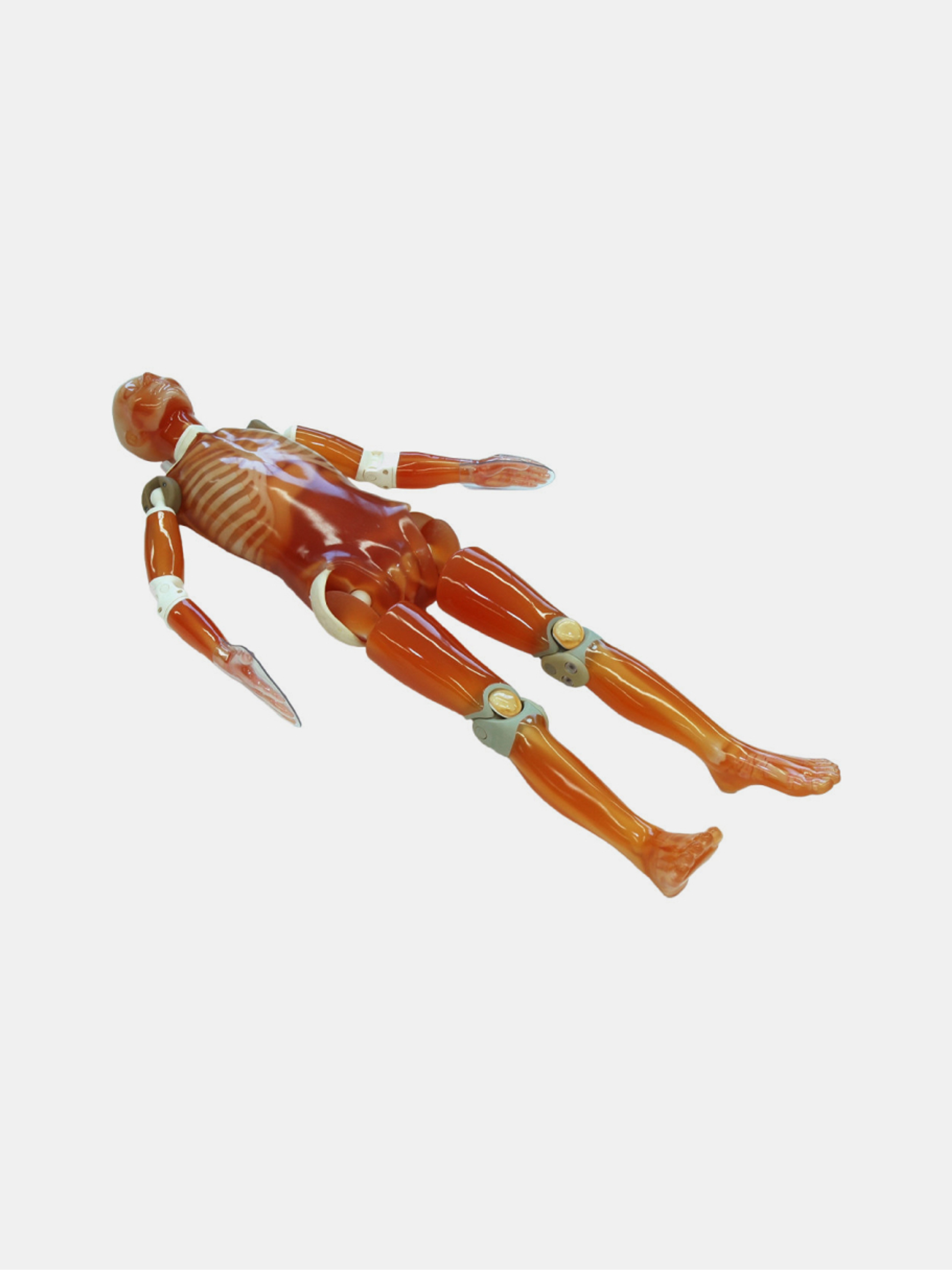
The CT Whole Body Phantom PBU-60 is a realistic anthropomorphic model designed for radiology training and CT imaging optimization
The CT Whole Body Phantom PH-2E is an adult anthropomorphic model with simulated pathologies for CT training and protocol optimization
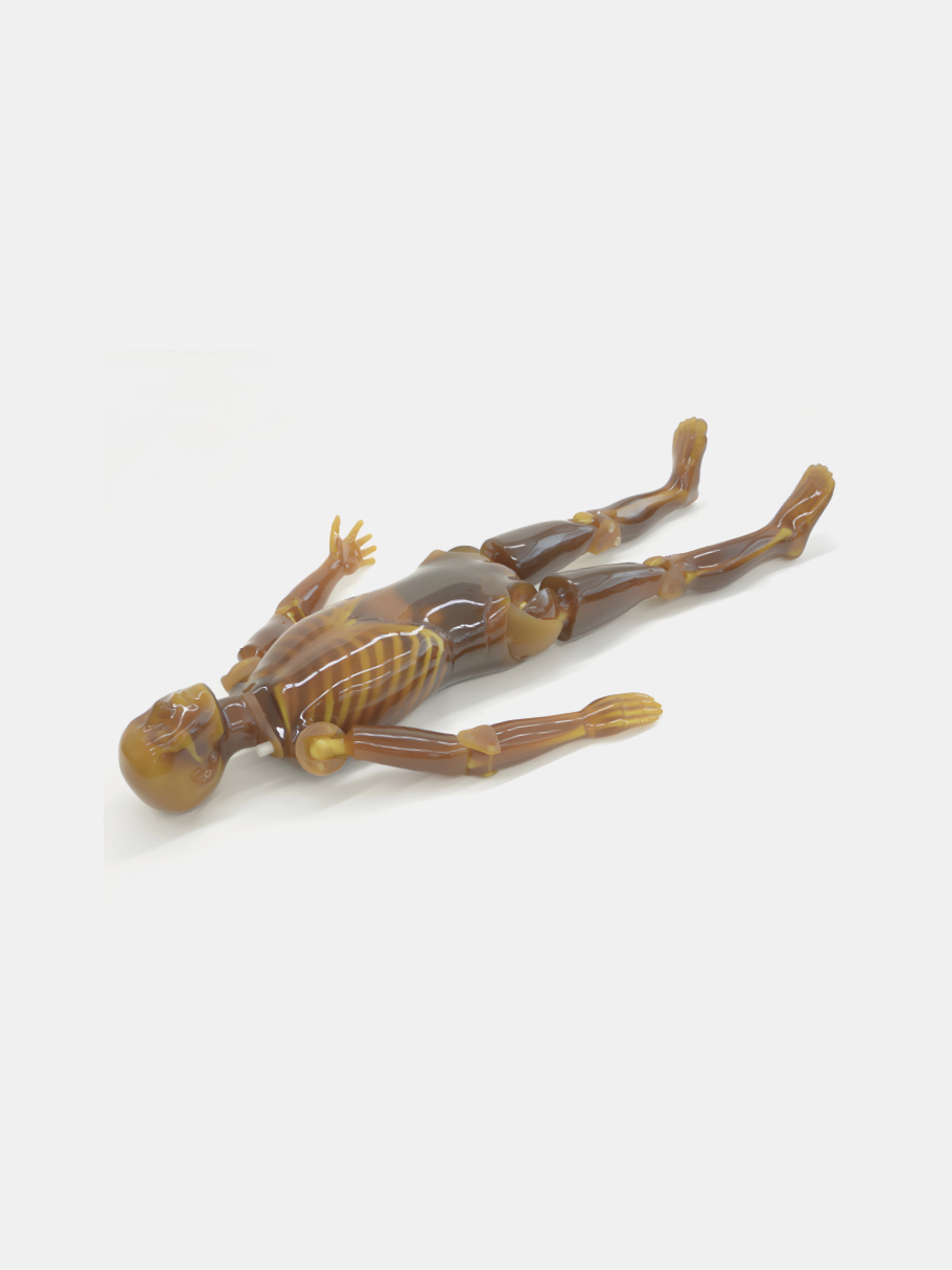
The PBU-90 Rugged Whole Body Phantom is designed for radiology and CT training with enhanced durability and easier handling
The PBU-50 Whole Body Phantom is an anthropomorphic disassemblable model designed for patient positioning training, plain X-ray, and basic CT
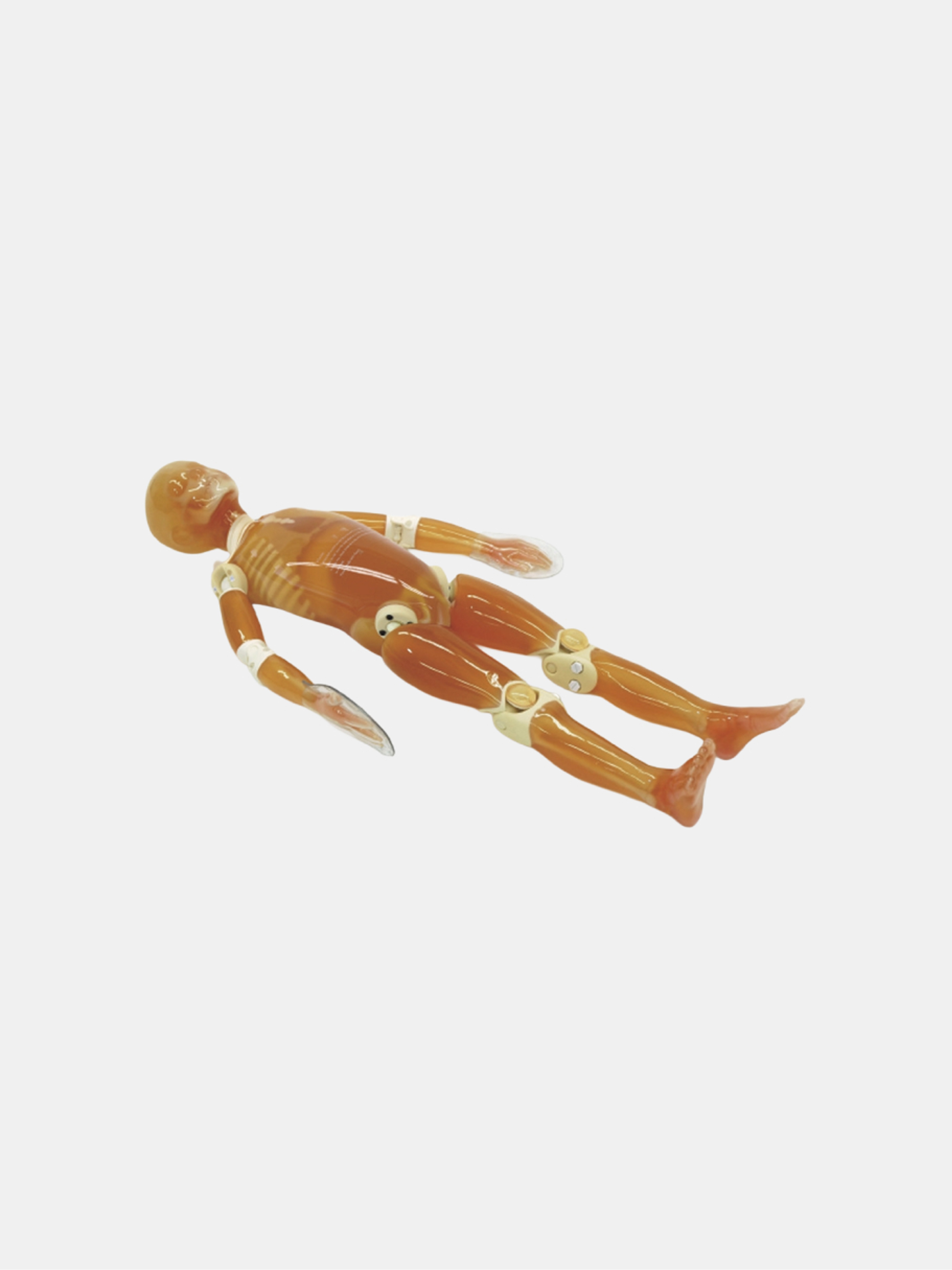
The PBU-70B pediatric phantom is designed for training in pediatric radiography, bone fracture detection, and awareness of child maltreatment
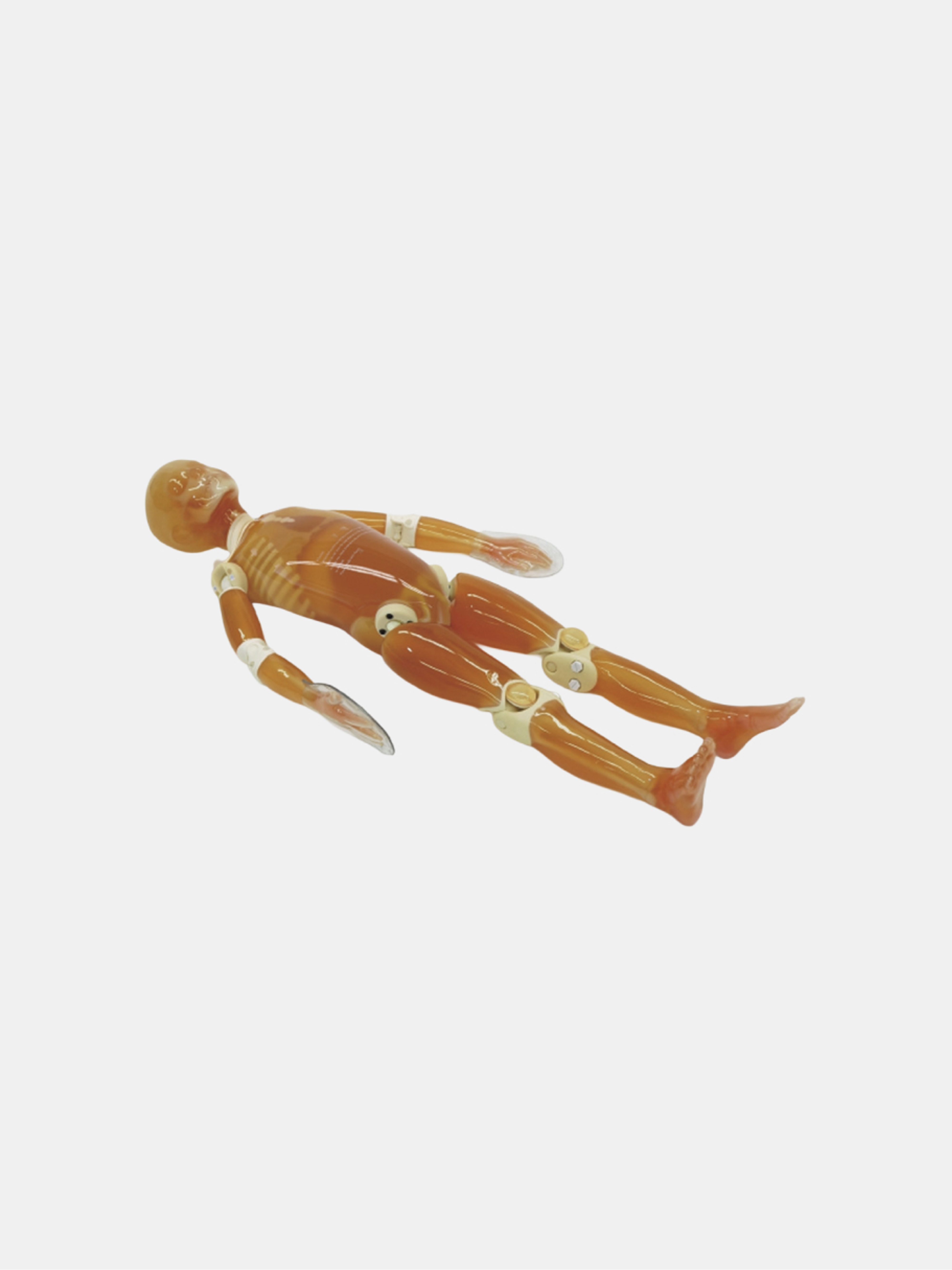
The PBU-70 Pediatric Whole Body Phantom represents a 5-year-old child, ideal for X-ray, CT, and basic patient positioning training
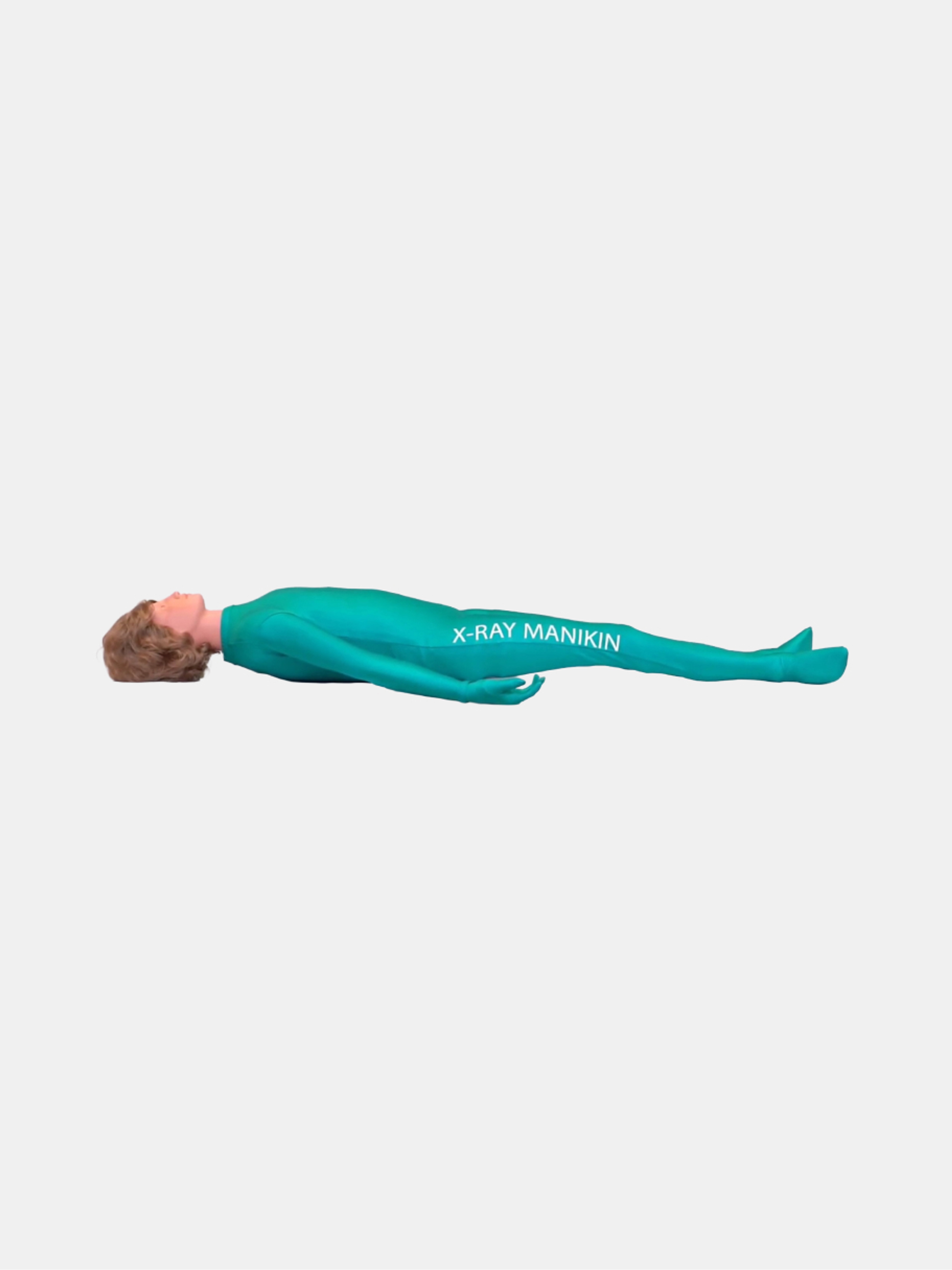
The Full Body X-Ray Phantom contains a natural human skeleton and provides realistic training for positioning and radiographic techniques
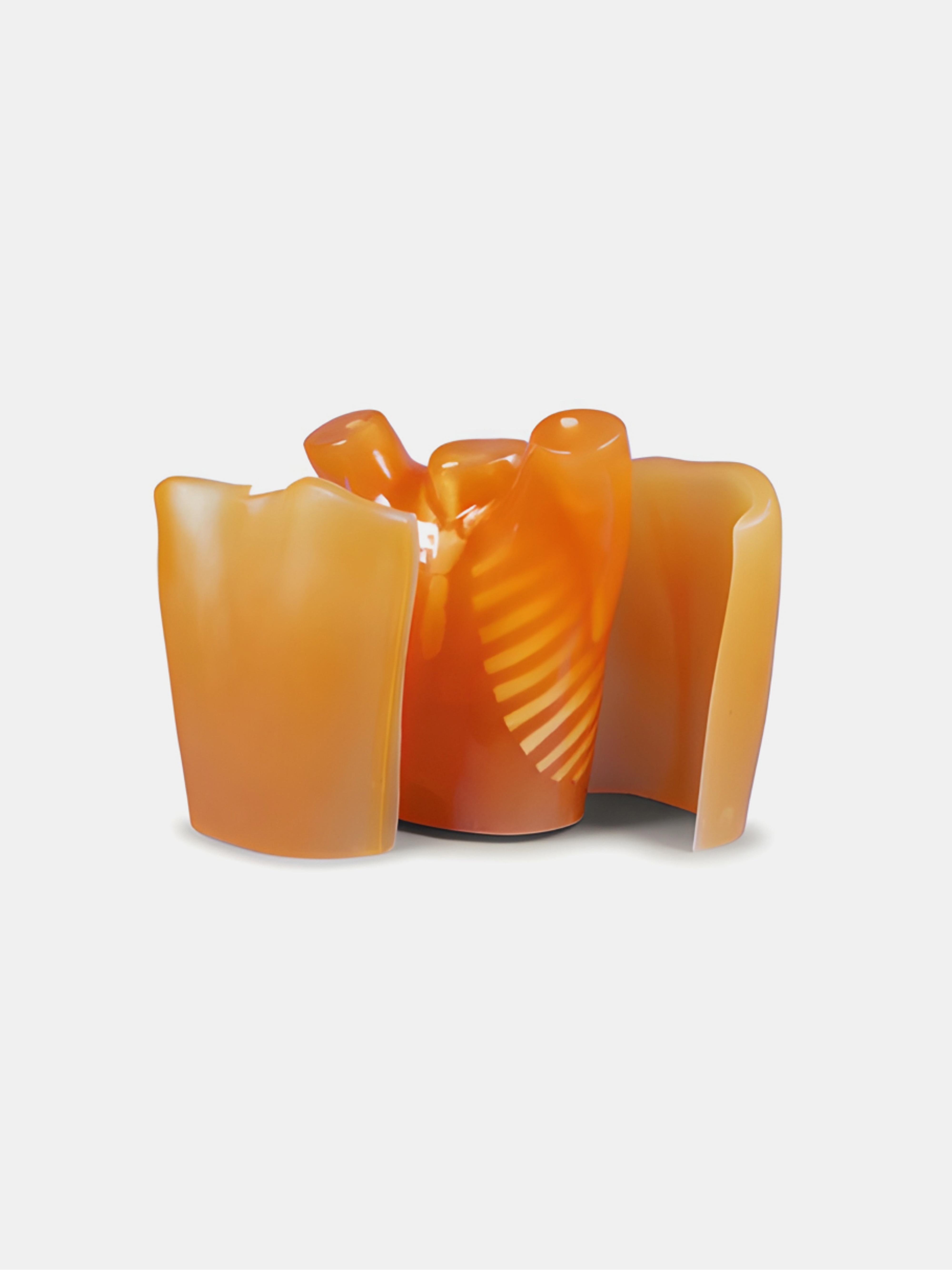
Lungman chest phantom in tissue-equivalent material for plain radiography and CT scanning, featuring simulated pulmonary vasculature for realistic protocols
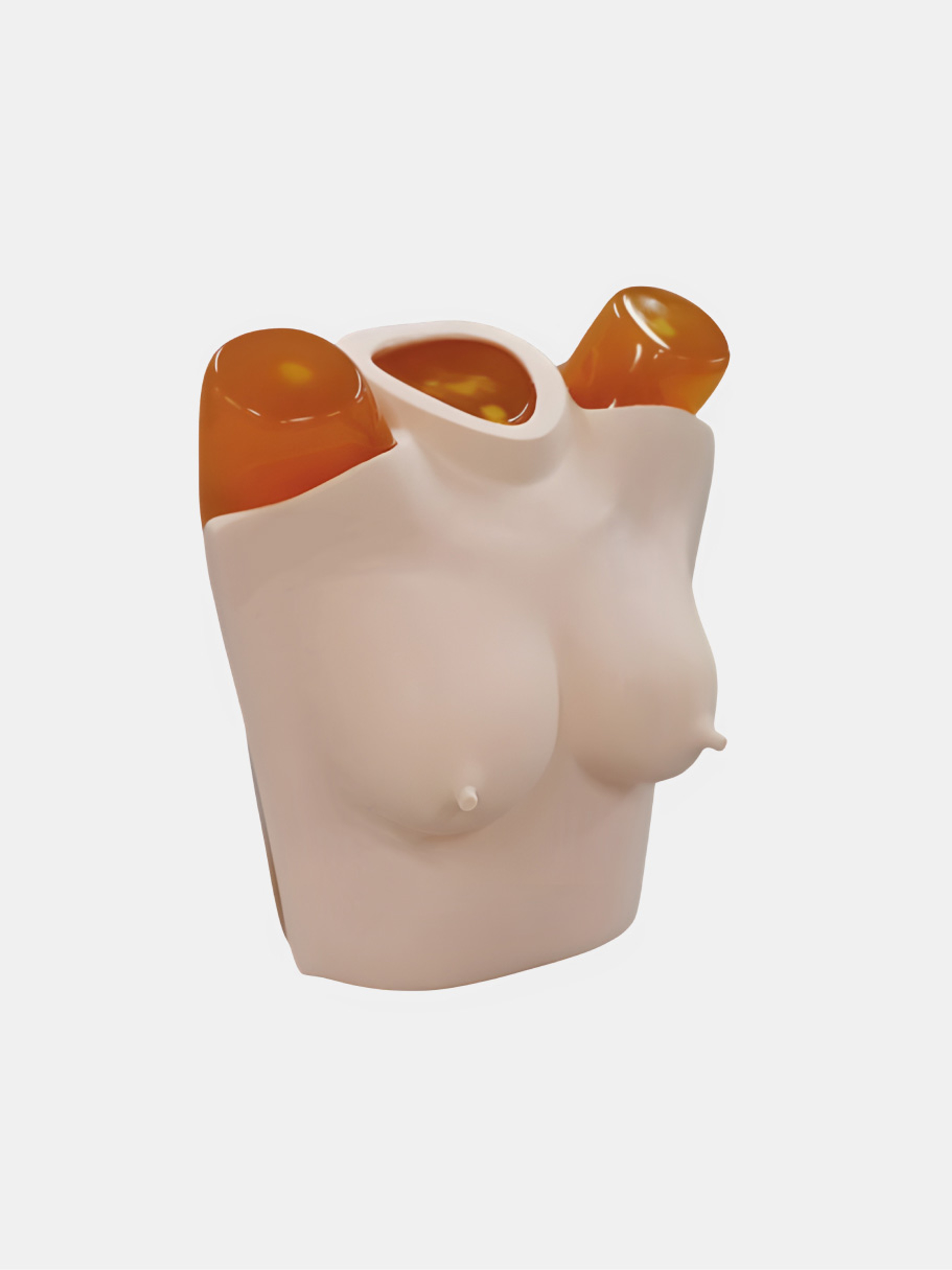
This plate simulates the absorption of breast in female chest X-ray, and the influence of nipple on image quality
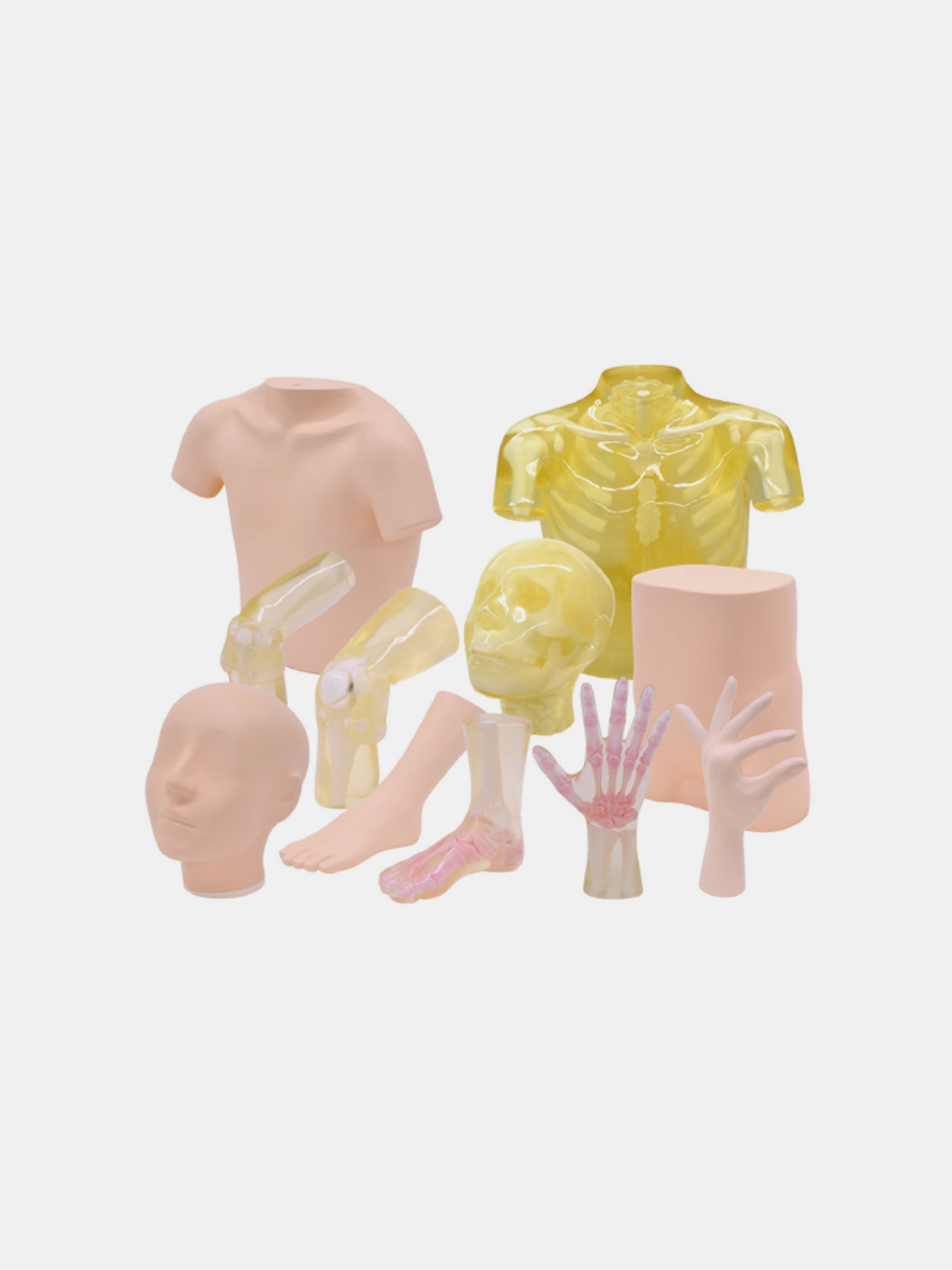
Sectional phantoms for X-ray imaging: head, thorax, pelvis and limbs, opaque or transparent models for radiographic positioning and training
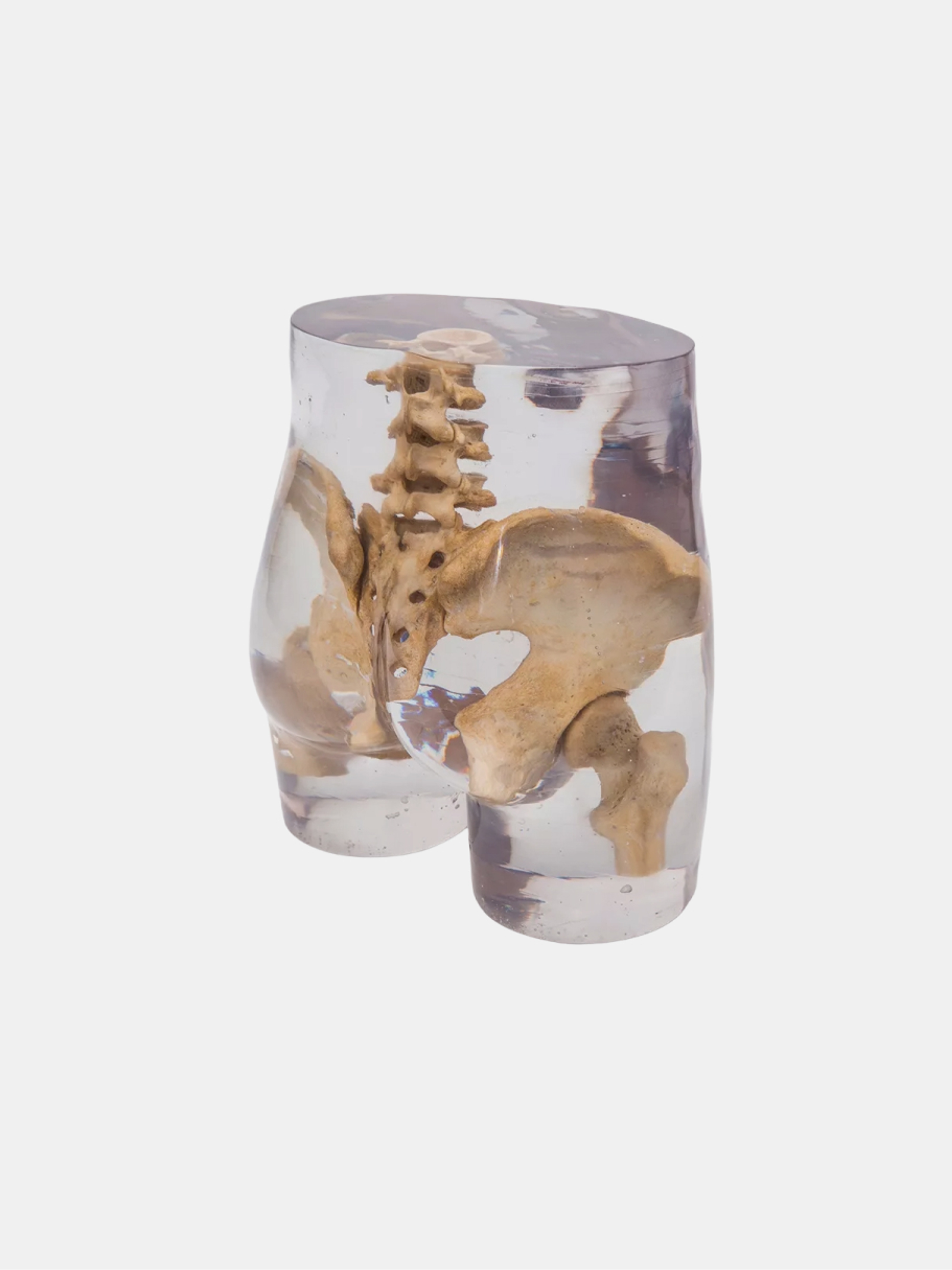
Transparent pelvis phantom designed for repeatable X-ray acquisitions in radiography without patient exposure
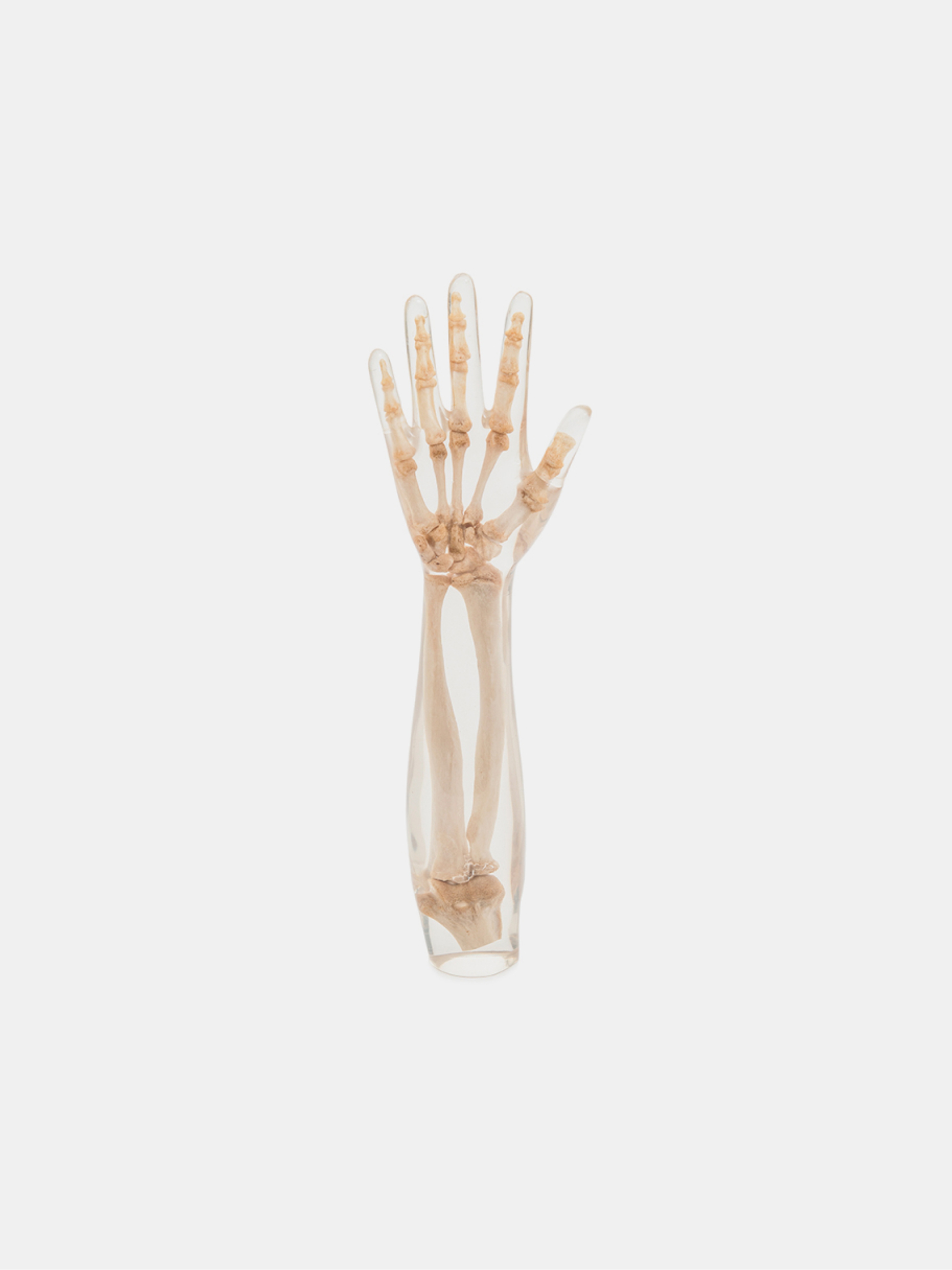
Forearm phantom with real human bones in transparent plastic, for radiology training and repeatable X-ray imaging without patient exposure
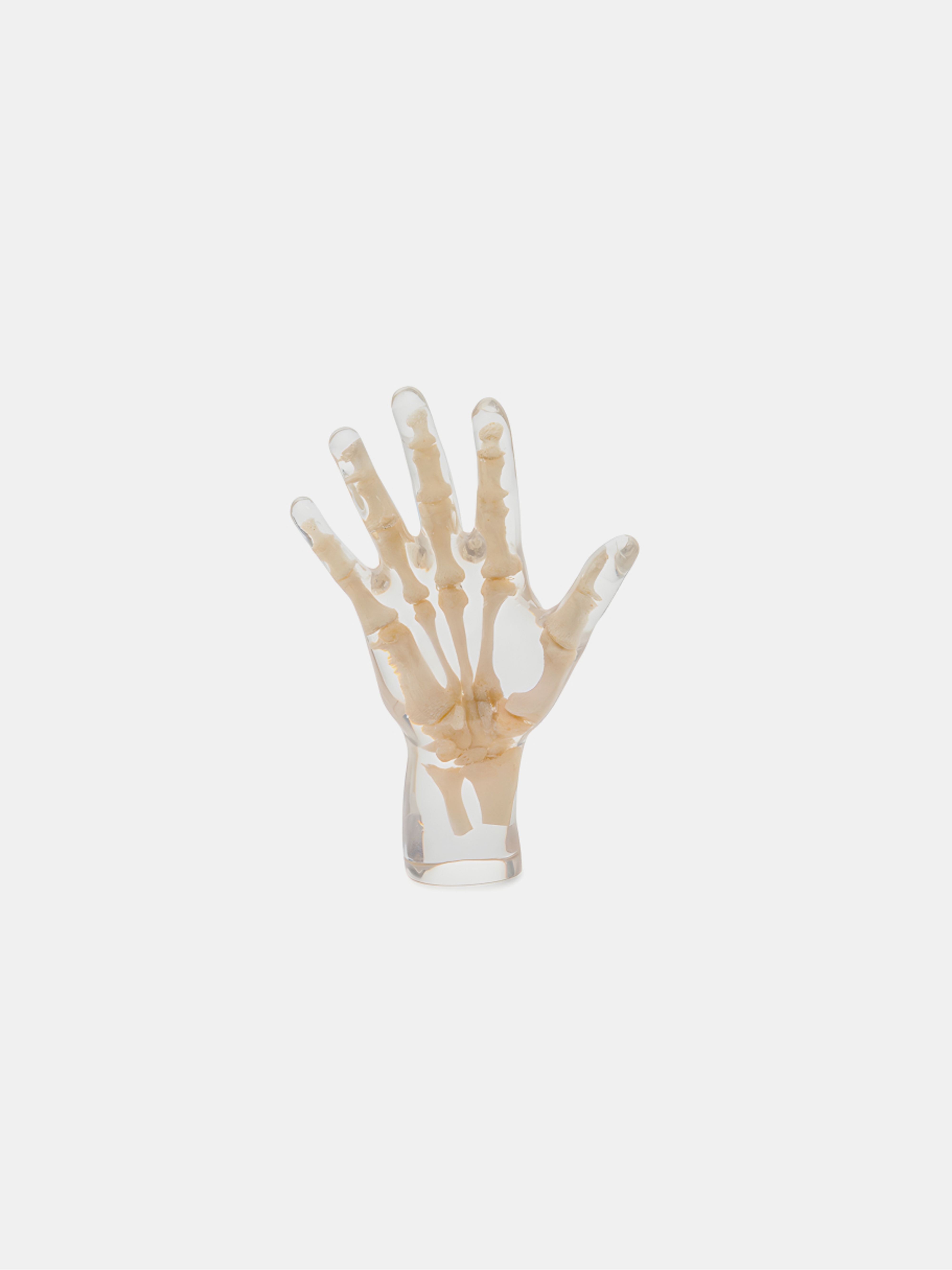
Hand phantom with real human bones in transparent plastic, for radiology training and repeatable X-ray imaging without patient exposure
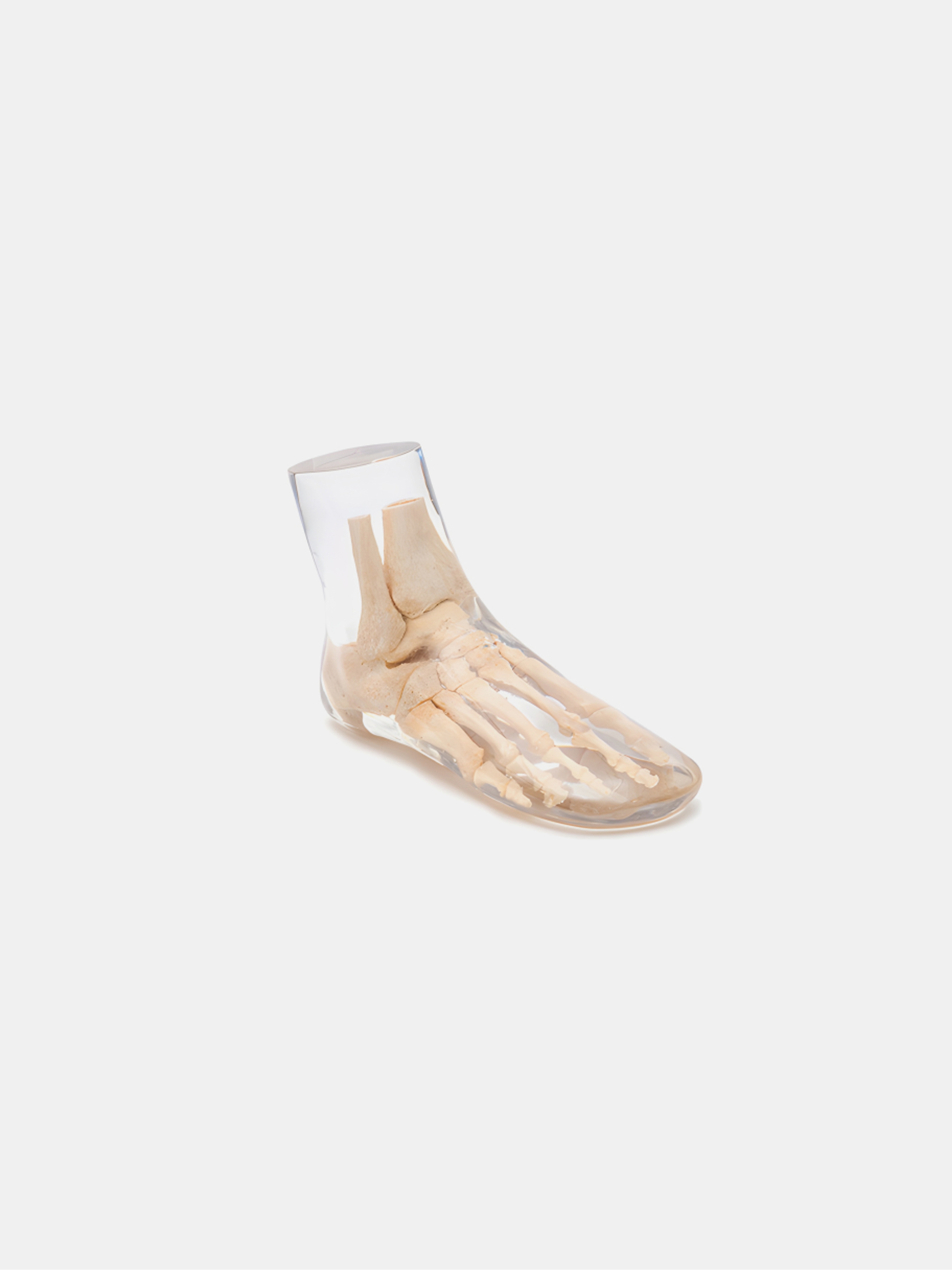
Foot phantom with real human bones in transparent plastic, for radiology training and repeatable X-ray imaging without patient exposure
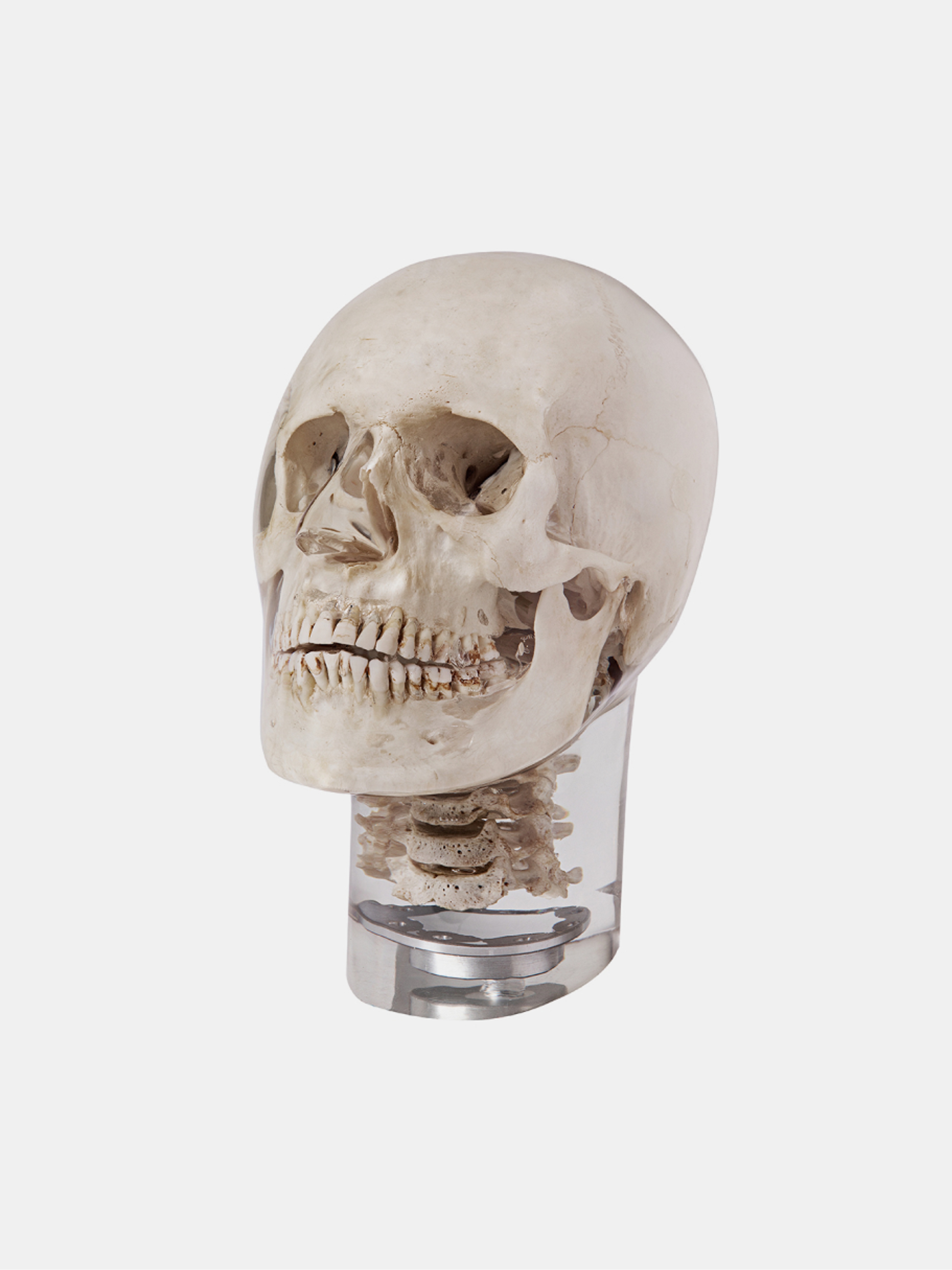
Human skull in natural bone embedded in plastic, jaws slightly open for dental panoramic X-ray imaging and radiological training
Radiology
Radiology is one of the founding disciplines of modern medical imaging. It relies on the use of X-rays to visualize the internal structures of the human body and to diagnose a wide range of pathologies.
Modern radiographic systems, whether direct digital (DR) or indirect (CR), provide high-resolution images that allow detailed examination of bones, lungs, the thorax, soft tissues and many other anatomical regions.
In addition to clinical equipment, anatomical radiology phantoms are used for training, research, protocol development and X-ray system demonstrations.
These models, representing whole bodies or realistic anatomical sections, faithfully reproduce the attenuation and scattering properties of human tissues. Some include simulated pathologies to evaluate lesion visibility and contrast under realistic clinical conditions.
Radiology therefore plays a key role in education, calibration and innovation in medical imaging, bridging clinical practice and applied physics in the service of accurate diagnosis.